 AIMALABS
A software-only AI platform for the blood smear.
Hardware-independent, microscope-agnostic, and built as assistive decision support — the specialist always confirms the result.
From slide to signed-off, in four steps.
Capture
Photograph or upload the stained smear straight from the microscope camera — no proprietary scanner required.
AI analysis
The model classifies cells and quantifies morphology across the blood-cell classes in under two minutes.
Structured output
White blood cell morphology, ICSH-aligned red cell morphology grading and platelet estimation, presented clearly.
Expert confirms
A pathologist or haematologist reviews and signs off. The AI supports the specialist; it never decides alone.
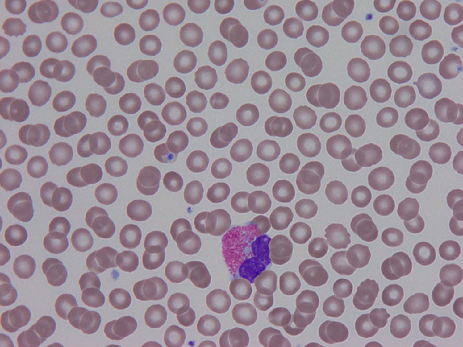
Peripheral blood smear (Wright–Giemsa) with an illustrative AIMALABS detection. Every result is confirmed by a qualified specialist.
From a field of cells to a structured readout.
On each smear, AIMALABS localises and classifies cells across the blood-cell classes, grades red-cell morphology to ICSH conventions, and estimates platelets — turning a microscope image into a consistent, reviewable result in under two minutes.
The advantages of losing the hardware.
No machine to buy
Deploys on the microscopes and cameras a lab already owns. No six-figure analyser, no rip-and-replace.
Fast, out of hours too
Roughly five times faster than manual review, available around the clock without a morphologist on site.
Consistent by design
The same standardised assessment on every smear, eliminating inter-observer variability.
Answers at the bedside
Rapid results support real-time decisions in acute settings where waiting isn’t an option.
Experts, better used
Frees trained scientists from routine screening to focus on complex cases and quality assurance.
Reaches everyone
Brings expert-level analysis to labs and settings that could never justify the hardware.
Built for the lab and the point of care.
AIMALABS runs as two variants of a single architecture: a full laboratory model for deployment in the lab, and a lightweight model for decentralised, point-of-care use — so the same standardised analysis follows the patient wherever the smear is taken.
Preliminary internal results on held-out data — not yet peer-reviewed or cleared.
See the platform on your workflow
Book a walkthrough and we’ll show AIMALABS running on smears like yours.
Book a demonstration →