 AIMALABS
Expert blood morphology, anytime, anywhere.
AIMALABS reads peripheral blood smears with AI on the microscopes and cameras labs already own — no new hardware, no capital cost. A standardised readout in under two minutes, ready for a specialist to confirm.
aíma · Greek for “blood” — the heart of everything we do.
The gold-standard test most patients never receive.
The peripheral blood smear is the gold standard supporting the diagnosis of leukaemias, anaemias and more. But manual review is slow, depends on scarce specialists, and the machines that automate it cost more than most labs can justify.
The test that finds what the count misses.
A blood count is automated and cheap — it answers how many cells? The smear is one glass slide read under a microscope, and it answers how do they look? That is where the diagnosis actually lives.
“How many cells?”
Automated · 5 billion a year. Fast and cheap — but blind to what the cells look like.
“How do they look?”
Manual · 10–20 minutes of expert time. Shape, size and structure of every cell — and 86% of the time it simply is not done.
How often the diagnosis comes late.
Leukaemia in England, measured across 12,770 cases. Behind every one of these sits the same bottleneck: 10–20 minutes of scarce expert time at a microscope, for every smear, by hand.
Leukaemia UK, “Every Diagnosis Counts” (2025), 12,770 cases in England · emergency-diagnosis rate vs all cancers: Leukaemia UK / NCRAS.
Any microscope becomes an expert haematologist.
AIMALABS is software only. It runs on the microscope and camera a laboratory already owns, returns a standardised read of the cells in under two minutes, and a morphologist confirms every result in one click. Nothing to buy, nothing to install on the bench.
Zero capital cost
Works on existing microscopes — no analyser, no scanner, no replacement of the bench.
An answer while the patient waits
The AI analysis takes under two minutes instead of 10–20 minutes of scarce expert time.
Assistive, never autonomous
A qualified morphologist confirms or overrides every result. AIMALABS supports the diagnosis; it never makes one.
One image in. A clinical readout out.
A software-only platform that works with standard microscopes and cameras — and always keeps a specialist in the loop.
Capture
Photograph or upload the stained smear from any microscope camera.
AI analysis
In under two minutes the model classifies cells and quantifies morphology.
Structured output
White blood cell morphology, ICSH-aligned red cell morphology grading and platelet estimation.
Expert confirms
A specialist reviews and signs off. AIMALABS assists — it never replaces them.
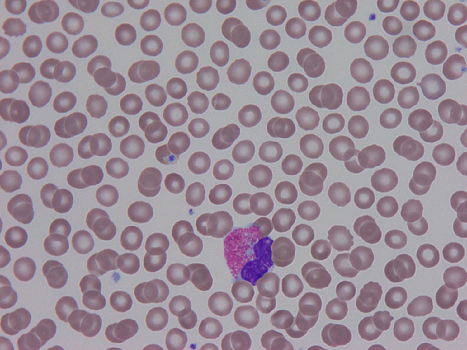
Real cells. Real-time analysis.
AIMALABS localises and classifies the cells on a stained peripheral smear — white cells, red-cell morphology and platelets — then hands a structured readout to your specialist to confirm.
Peripheral blood smear (Wright–Giemsa). Coral overlay: an illustrative AIMALABS cell detection.
Two ways to read a blood film.
For the patient: an answer before they leave — not a letter weeks later.
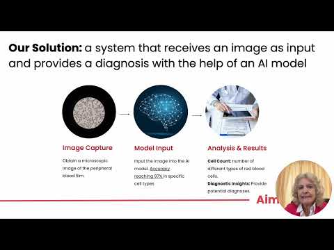
Watch the demo, the 2-minute video and the pitch.
Three short videos on what AIMALABS does and why it matters.

What this means for a laboratory.
Scarce experts, better used
Routine screening is handled first-pass, freeing trained scientists for complex cases and quality assurance.
Cover on every shift
The same standardised read at 3am as at 3pm — no waiting for a morphologist to come on shift.
Wherever the smear is taken
Software-only delivery brings expert-level morphology to labs and settings that could never justify the hardware.
Ready to see AIMALABS on your own smears?
Whether you’re a laboratory, a hospital, an industry partner or an investor — we’d welcome a short conversation.
Book a demonstration →